 Built-in viewer
A DICOM viewer for the people who keep imaging running.
For looking at studies from PACS, archives and cloud stores, finding out why one looks wrong, hanging mammograms the way they're read, and learning the anatomy with free organ outlines. In the browser, built on Cornerstone3D, with nothing to install. Non-diagnostic; not FDA cleared.
Looking for the free viewer that goes on patient discs? See the portable viewer
For PACS admins & radiology IT
New in 7.0What happens when a PACS admin builds a viewer
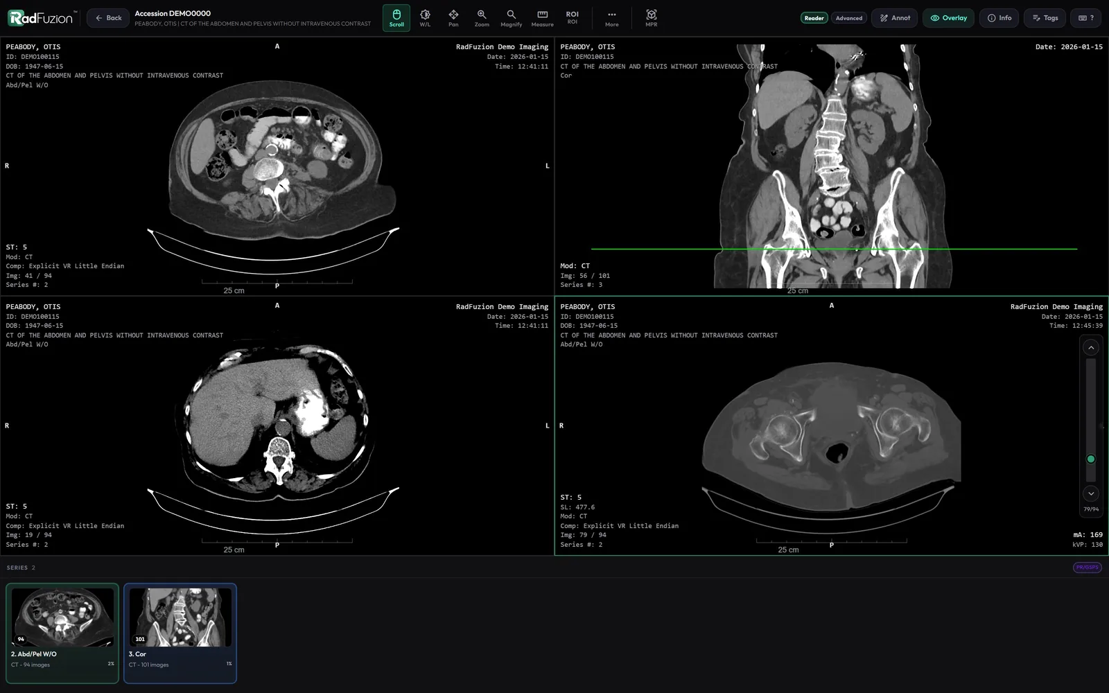
When a study looks wrong on the PACS, the first question is always the same: is the problem in the images, or in the PACS? Most viewers are built to read studies. This one is also built to answer that question.
Open Inspect beside the images and it reads every header in the series or the whole exam and tells you, in plain words, what's wrong: slices missing and where, an image tilted differently from the rest, another patient's image in the series, two images with one UID, a window that changes on every slice. Click an image number to see it. Click a tag to find it.
“I've been the PACS admin, the network engineer and the HL7 engineer for a teleradiology practice since 2010. These are the checks I've done by hand for years. Now they're one click in the viewer.”

What's in Inspect
Check
Reads every header in the series or the exam: missing or uneven slices, mixed orientations, gantry tilt, several stacks in one series, repeated Instance Numbers, duplicate UIDs, another patient or study mixed in, fewer images than the scanner made.
Compare
Two images, or every image in a series, side by side: only the tags that differ, with which images have each value. Tags that change per image anyway are set aside.
Validate
The rules a strict PACS enforces: required tags, dates, times and UIDs in the right form, value counts, File Meta that matches the dataset, pixel data that isn’t cut off.
Display
Every header value that decides how the image looks, step by step: bits, rescale, each header window, VOI LUT, MONOCHROME1, presentation state, overlays, shutters. Switch one at a time to see which one a PACS treats differently.
Changes
What was changed in the image after it was made, from its Original Attributes record: before and now, by which system, when. Plus RadFuzion’s own change log for the exam.
Trail
Where the exam came from (AE title, address, time), which rules it matched, where it went and how far it got, and every event along the way.
Export
The original file, a de-identified copy for a vendor ticket (it warns when the pixels may hold names), or the tags as text or JSON. Every export is in the Access Log.
DICOM tag search
Search an image’s tags by number in any form (0018,0050 or just 0019), by name, or by value. Sequences open into their items; private and empty tags are listed too.
Overlays & shutters
Overlay planes and display shutters are drawn the way a PACS draws them, and can be turned off to see what’s underneath.
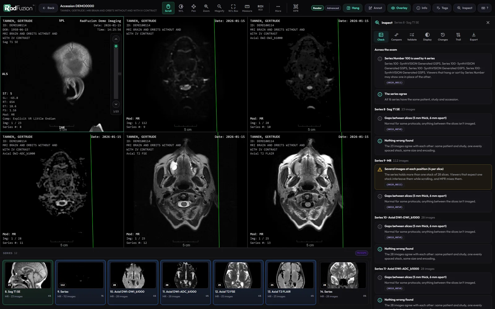
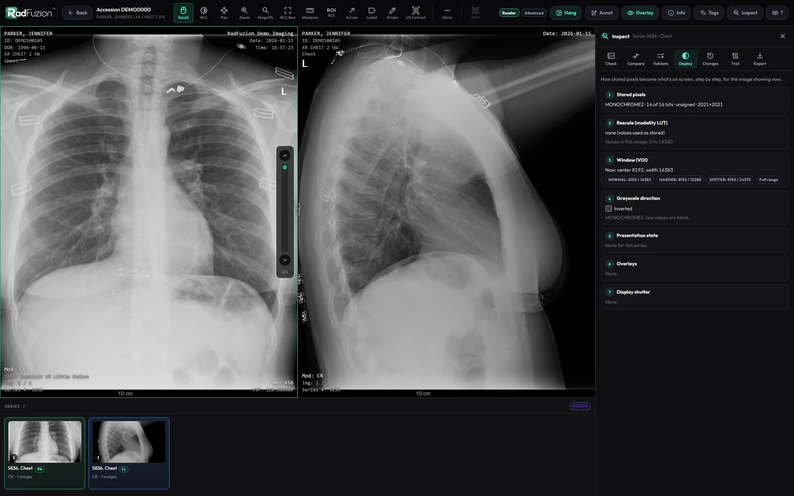
Inspect is part of the viewer in every RadFuzion license. It needs the Tag Editor view permission, works on exams RadFuzion received, the image cache, and Local Storage, and never changes the images. The viewer is non-diagnostic and not FDA cleared for primary diagnostic interpretation: Inspect is for troubleshooting the images, not reading them.
Everything else RadFuzion does for PACS adminsMammography
New in 7.0Mammograms hung the way they're read
Right and left back to back, each chest wall against the edge, zoomed to the breast instead of the black around it. The corner text moves to the outside, off the image. Tomosynthesis plays every view together, and the CAD marks land on the right view even when the CAD report points at images that aren't in the exam.
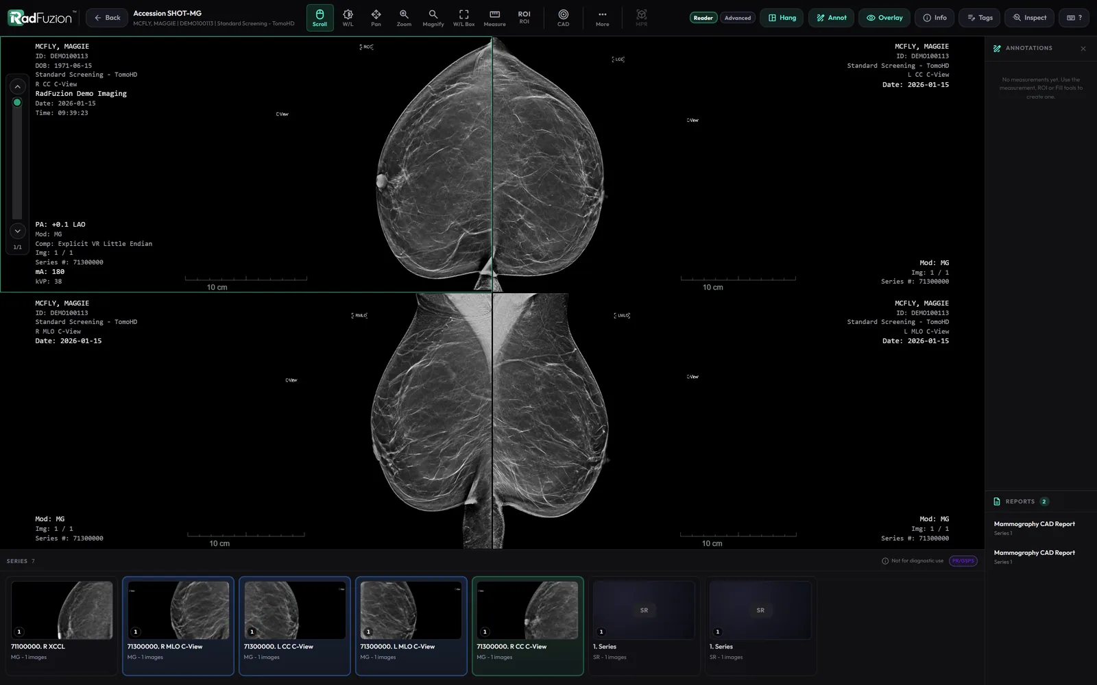
Edge to edge, filled
Zoomed to the breast. Hold Zoom for the whole image or 1:1 pixels, or press Alt+0. Reset puts it back.
Tomo in step
Every view plays from slice 1. Views with fewer slices move slower, so they all reach the end together. Scroll one and the rest follow.
CAD on every view
Marks on the 2D and on each tomosynthesis slice, with the findings listed by view. One button, remembered.
Cine at your speed
Hold Cine for 10 to 200 frames a second, or Auto. Scroll to stop on the slice you want.
Organ outlines
Free add-onPoint at it, and it tells you what it is
Everyone who supports imaging has looked at a CT and wondered which gray shape is the spleen. Install the free organ-outline add-on, right-click a CT or MR series, and pick Organs. A minute or a few later every structure it found is outlined in color. Point at one with any tool and its name shows beside the pointer.
It runs TotalSegmentator, the open-source model from University Hospital Basel. No GPU, no problem: it runs on your server's own processor and takes a couple of minutes. If there's an Apple silicon Mac or a Linux machine with an NVIDIA card on your network, a processing worker can hand the job to its GPU instead, at full resolution, with the patient's details left off the images. Nothing leaves your network. It's a learning tool, not a diagnosis.

One click to install
Settings › Add-ons downloads it once (about 3 GB) from where each part is published. Nothing runs until someone asks.
Your CPU, low priority
More than 100 structures on CT, about 50 on MR, on the server’s processor. Routing comes first; one series at a time.
Groups & spine labels
Key organs first, then the skeleton, vessels, muscles or everything. On CT it can label each vertebra, C1 to S1.
Kept & exported
The outlines stay with the exam while it’s on the server. Volumes as CSV, the labels as NIfTI or a DICOM SEG.
Free with every RadFuzion license. It uses only TotalSegmentator's open (Apache-2.0) tasks, with its usage statistics turned off. TotalSegmentator is the work of Wasserthal et al., University Hospital Basel, and isn't affiliated with RadFuzion. Not for diagnosis.
Portable viewer
Free with RadBurnA different viewer goes on patient discs
Discs burned with RadBurn carry a small viewer of their own, so patients and referring providers can open the images with nothing to install. It has its own page, with a live demo you can try.
See the portable viewerarrow_forward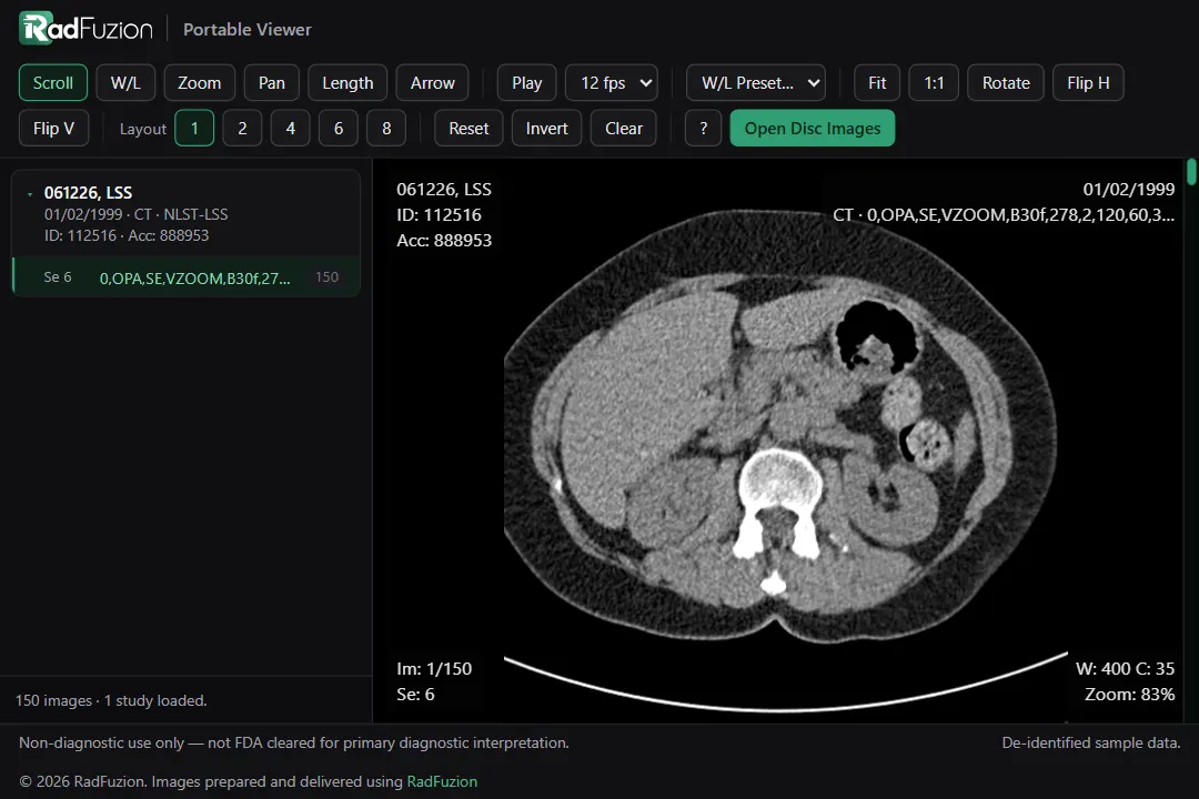
What the viewer does
MPR, MIP & 3D
Axial, coronal, and sagittal MPR with MIP, MinIP, and thick slab, plus 3D volume rendering with tissue presets.
Window / level
One-click presets for CT, MR, PET, ultrasound, and more, plus interactive windowing.
Measurements & annotations
Length, angle, Cobb, ellipse, freehand, spline, and livewire ROIs, probe/HU, arrows, and labels.
Layouts & linked scrolling
1 to 10 viewports, linked scrolling, and localizer lines across series.
Hanging protocols
Exams open by modality: mammograms back to back, X-rays one view each, CT in its three planes, MR by sequence, scanned paperwork last.
ECGs, video & AI results
12-lead ECGs on their grid, MP4 video, and SEG, RT structure set and SR results drawn on the images.
Cine & tomosynthesis
Cine at 10 to 200 frames a second, or Auto, and tomosynthesis with every view playing in step.
Reader and Advanced modes
A lean Reader mode for fast reading, or the full toolset, with keyboard shortcuts.
Readable SR reports
Structured reports show as text in the sidebar, and Send ORU turns a final report into HL7.
Key images & spine labels
Mark key images and label vertebrae, then jump every viewport to a level.
Undo & redo
Ctrl+Z and Ctrl+Y for measurements drawn, moved or deleted, Clear All included, and segmentation.
Fill
Click a region to outline it by its values, drag to grow it, and read its area, mean and range (HU on CT).
Image registration
A free add-on lines up two series, rigid or affine, with before, after and checkerboard views to judge the fit.
Inspect, for PACS admins
Check a series or exam, compare and validate tags, see every display setting, and trace where the exam came from and went.
Compressed images
JPEG 2000, HTJ2K, and color images display directly in the browser.
See the viewer on your studies.
Request a walkthrough and we’ll show it running against your environment.
mailRequest a demo